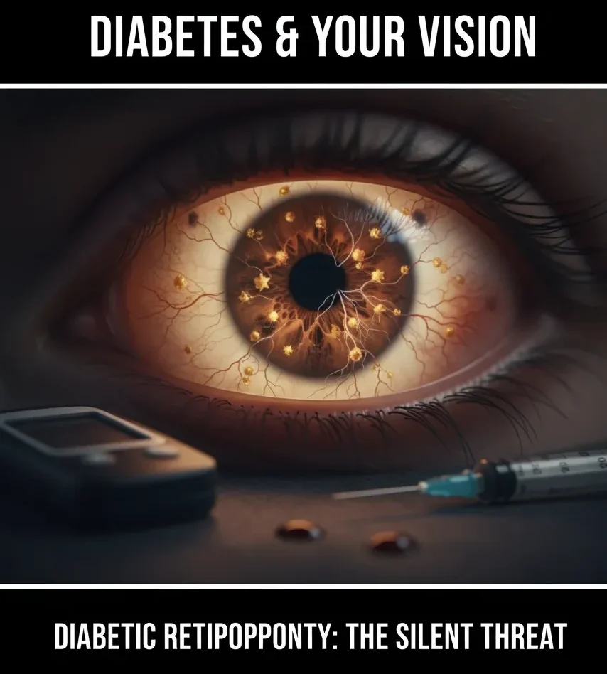
Diabetes doesn’t just affect your blood sugar; it can also silently damage your eyesight. Over time, high blood sugar levels can harm the tiny blood vessels in the retina, leading to diabetic retinopathy, one of the leading causes of vision loss among adults.
Fortunately, with regular diabetic eye care in Hoshiarpur and early diagnosis, this condition can be managed effectively, helping you maintain clear vision for life.
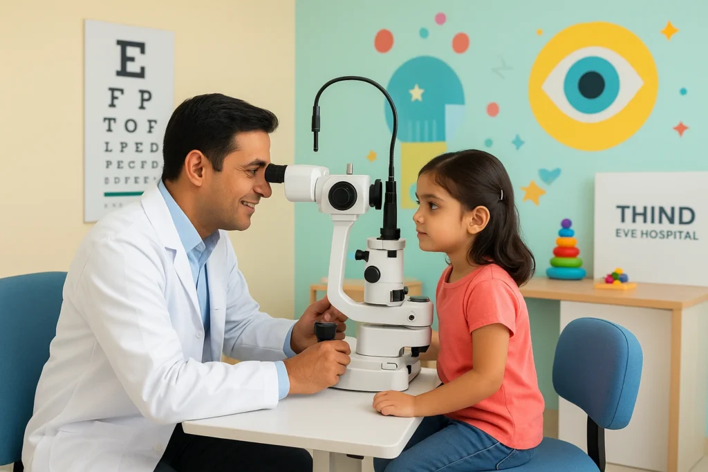
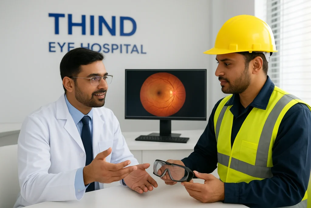
At Thind Eye Hospital, a NABH-accredited super-speciality centre, patients have access to advanced diagnostics, state-of-the-art retinal imaging, and leading retina specialists near you. This combination of expertise and technology ensures timely detection and personalised diabetic eye management for every patient.
Why Diabetic Eye Care Is Essential
Diabetic eye diseases often progress quietly, showing few or no symptoms in the early stages. By the time vision blurs or dark spots appear, irreversible damage may already have occurred.
Regular Diabetic Eye Exams Help To:
- Detect diabetic retinopathy early
- Monitor retinal blood vessels and macular health
- Prevent severe complications like retinal detachment or blindness
- Guide timely laser or injection-based treatments
Experts recommend that every person with diabetes undergo a comprehensive eye exam at least once a year, even if their vision seems normal.
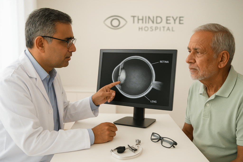
Understanding Diabetic Retinopathy
Diabetic retinopathy is a complication of diabetes that affects the retina, the light-sensitive tissue at the back of the eye.
Types of Diabetic Retinopathy:
- Non-Proliferative Diabetic Retinopathy (NPDR):
- Early stage
- Tiny blood vessels leak fluid or blood, causing swelling in the retina.
- Non-Proliferative Diabetic Retinopathy (NPDR):
- Proliferative Diabetic Retinopathy (PDR):
- Advanced stage
- New abnormal blood vessels form, increasing the risk of retinal bleeding and vision loss.
- Proliferative Diabetic Retinopathy (PDR):
If not treated promptly, both can progress to diabetic macular oedema (DME), swelling of the central retina that causes blurred or distorted vision.
Thind Eye Hospital’s specialists are highly experienced in diagnosing and treating all stages of diabetic retinopathy in Hoshiarpur using world-class technology.
Warning Signs You Shouldn’t Ignore
Even if you don’t notice changes in your eyesight, diabetes can still damage your retina. Seek medical attention immediately if you experience:
- Blurred or fluctuating vision
- Dark or empty spots in your sight
- Difficulty seeing at night
- Frequent changes in your glasses prescription
- Eye floaters or flashes of light
Early evaluation by a retina specialist near you can save your vision and prevent long-term complications.
Comprehensive Diabetic Eye Care at Thind Eye Hospital
Thind Eye Hospital offers a complete range of diabetic eye care in Hoshiarpur, focusing on early detection, preventive management, and advanced treatment.
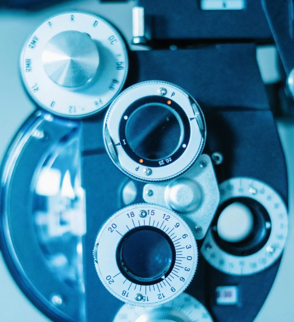
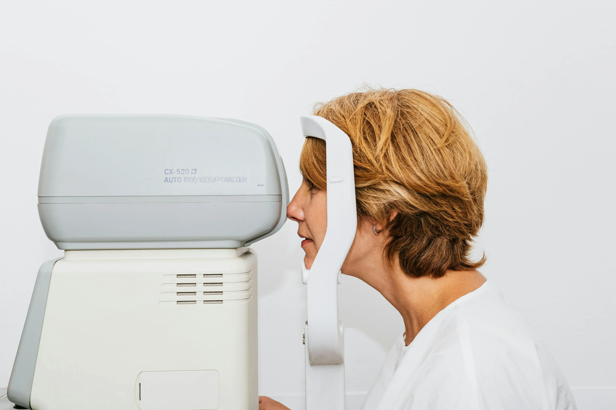
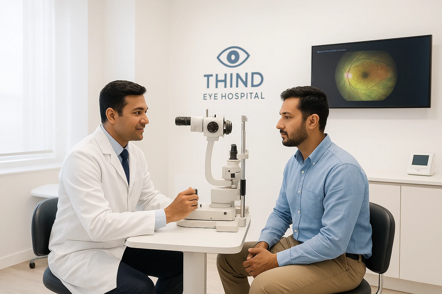
1. Retinal Screening & Imaging
Using advanced diagnostic tools such as:
- Optical Coherence Tomography (OCT): Provides 3D images of retinal layers for detailed analysis.
- Fundus Fluorescein Angiography (FFA): Detects abnormal or leaking blood vessels.
- Digital Fundus Photography: Helps monitor diabetic changes over time.
- Digital Fundus Photography: Helps monitor diabetic changes over time.
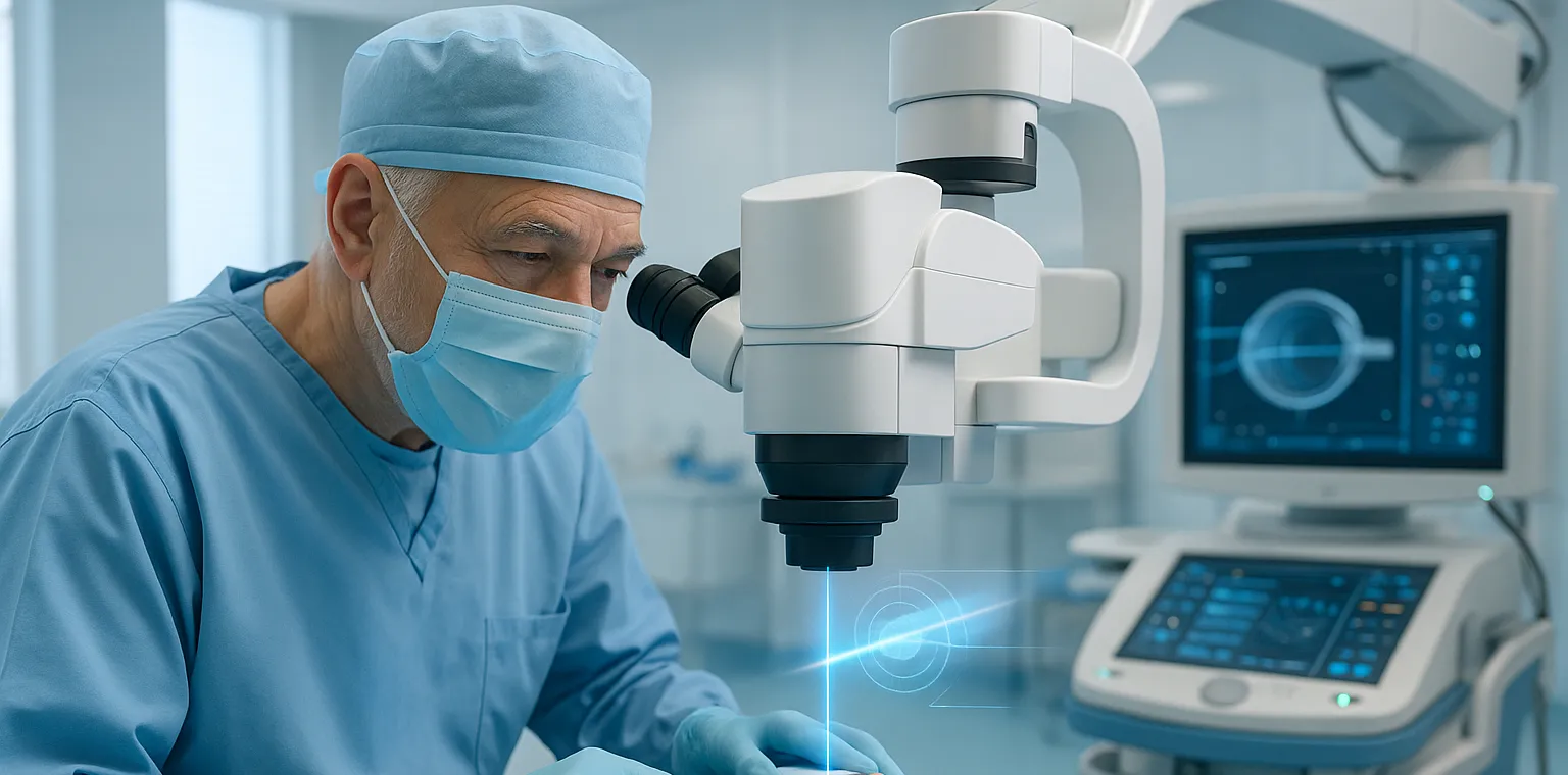
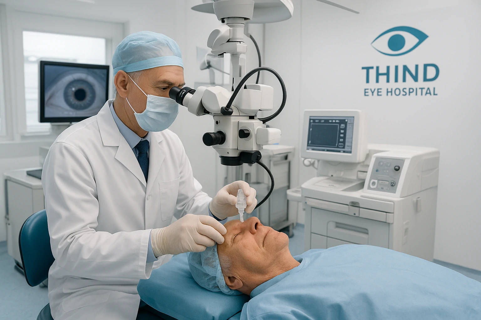
2. Laser Treatment (Photocoagulation): Laser therapy seals leaking vessels and prevents further retinal damage, effectively controlling diabetic retinopathy in Hoshiarpur.
3. Anti-VEGF Injections: Used for macular oedema, these injections reduce fluid leakage and swelling in the retina, restoring vision quality.
4. Vitrectomy Surgery: In advanced cases, minimally invasive vitreo-retinal surgery helps remove blood or scar tissue from inside the eye to restore sight.
5. Ongoing Monitoring & Counselling: Thind Eye Hospital’s diabetic eye program includes regular checkups, lifestyle guidance, and coordination with your diabetologist for comprehensive care.
Why Choose Thind Eye Hospital for Diabetic Eye Care in Hoshiarpur
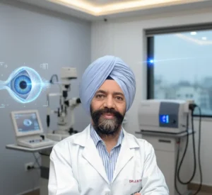
1. Expert Retina Specialists: Led by Dr. Kanwaljeet H. Madan and Dr. Sangeet Mittal, both renowned for their excellence in vitreo-retinal care, the hospital provides unmatched expertise for complex diabetic eye conditions.
2. Advanced Diagnostic Technology: The hospital uses cutting-edge equipment like OCT, FFA, and laser platforms for accurate and early detection, ensuring no retinal detail goes unnoticed.
3. NABH Accreditation & Proven Safety: As a NABH-accredited eye hospital, Thind Eye Hospital guarantees the highest quality of hygiene, safety, and clinical precision.
4. Comprehensive Eye Care Under One Roof: From diabetic screening and retina management to cataract and LASIK, all treatments are available locally, so patients no longer need to travel to metro cities.
5. Compassionate & Preventive Approach: Thind Eye Hospital focuses on education and early screening, empowering diabetic patients to take control of their vision health.
How to Protect Your Eyes from Diabetic Damage
Simple Preventive Steps:
- Keep your blood sugar, blood pressure, and cholesterol under control.
- Have regular eye exams every 6–12 months.
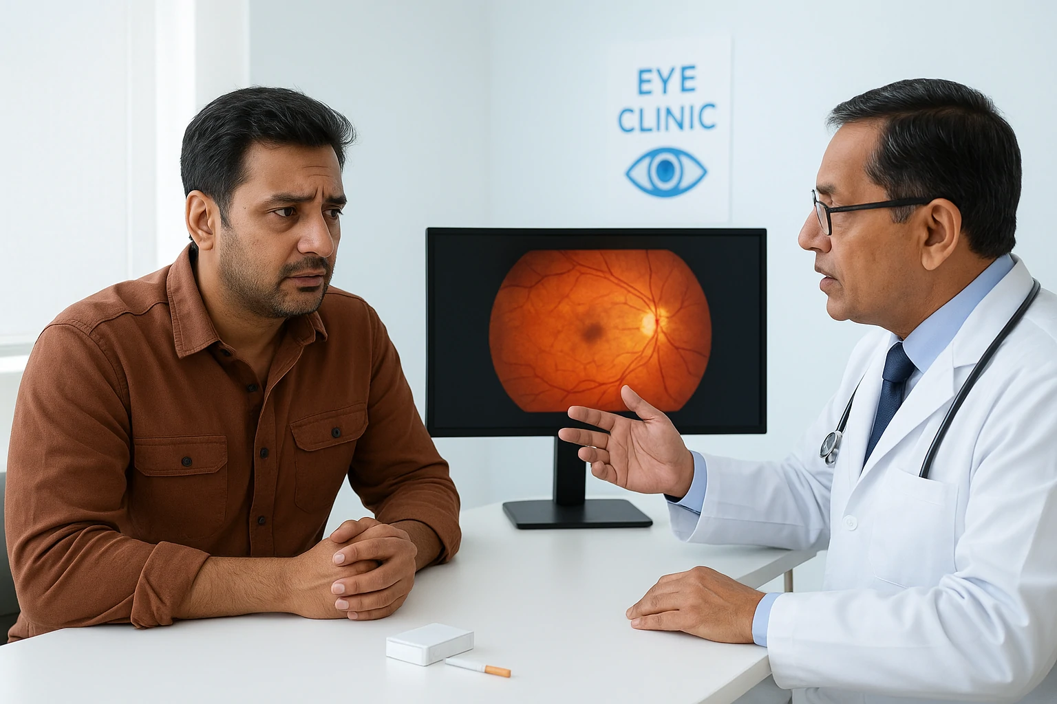
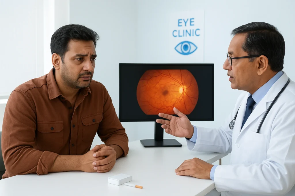
- Quit smoking, it worsens blood vessel damage.
- Eat antioxidant-rich foods (green vegetables, fish, citrus fruits).
- Stay active and maintain a healthy weight.
Early intervention can make the difference between lifelong vision and irreversible loss.
Patient Story: From Blurred Vision to Bright Clarity
“I had diabetes for 10 years, but ignored my eye health. During my visit to Thind Eye Hospital, I was diagnosed with early diabetic retinopathy. Thanks to timely laser treatment, my vision is back to normal. The doctors were caring and explained everything in detail.”
— Sukhdev Singh, Hoshiarpur
This story highlights the importance of regular diabetic eye care in Hoshiarpur and the impact of early detection.
Conclusion
Diabetes may be lifelong, but vision loss doesn’t have to be. With early detection and expert management at Thind Eye Hospital, patients can protect their eyes from diabetic complications.
As the leading centre for diabetic eye care in Hoshiarpur, equipped with advanced imaging and experienced retina specialists, Thind Eye Hospital ensures every patient receives the care they deserve, before it’s too late.
Book your comprehensive diabetic eye screening today and take the first step toward lifelong vision health.