Cataract and Your Eyes

Cataract and Your Eyes

A cataract is a cloudiness or opacity of the eye’s natural clear lens. It obstructs the passage of light to the retina (parda) of the eye and impairs vision. When the lens develops cloudiness to the point that it impairs vision, it is called cataract. It is like looking through a frosted glass.

How is cataract diagnosed?
The ophthalmologist examines the rest of the eye so as to predict the visual results of the surgery. The pressure of eye is recorded so as to rule out glaucoma. If possible, the retina is also examined, otherwise ultrasound scanning is performed.

What are the symptoms?
Progressive and painless blurring of vision is the commonest symptom. The blurring of vision may be more marked in bright light. Brightness and contrast of the image decreases in early cataract. Some individuals experience glare in night driving. Colors may become less district. Later on the vision deteriorates and interferes in day to day activities. Seeing two or more moons poyopial at night also suggests cataract. Pain, itching, redness or watering are not symptoms of cataract. Cataract usually develops in both the eyes, but may progress at different rates.

Is there a cure?
Although research is currently underway, no preventive measures are known for cataract that develops with age. No diet, drugs or medicine have been proven to delay or cure the cataract. The only treatment is surgical. Cataract surgery is a safe surgical procedure, with the appropriate lens implant, has restored sight for millions.

Phacoemulsificatlon or Phaco


Phacomulsification                                                            Conventional Surgery
1.2 mm to 1.8 mm long Incision                                             7 mm to 9 mm long Incision

It is a no stitch small incision cataract surgery. IOL is implanted and the surgery is completed without any stitch. The Incision is only 1.8mm – 2.2mm long.

A computer controlled ‘Phaco’ machine with tiny ultrasonic probe is used to remove cataract. Phaco uses ultrasound energy to liquify lens and suck it out with a titanium probe, vibrating forty thousand times a second. The lens is implanted through the smallest possible opening in the eye. The minute (2.2mm or smaller) incision is self sealed and needs no suture or stitch to close it. The patient is back to normal activities within a few days, including attending office or even doing heavy manual work. Phacoemulsificatlon was started for the first time in Jalandhar at THIND EYE HOSPITAL. At present this technique is used for all cases undergoing Cataract surgery at our hospital. To retain the maximum benefit of the miniature 2.2mm or smallest incision, a foldable IOL is used. The foldable IOL made of transparent silicone or acrylic polymer to be inserted inside the eye in a fine tube and it unfolds to It’s full size once inside the eye.


FAQs

What should I do right after cataract surgery?
Plan to spend the day resting quietly when you return home after surgery. For a day or two, avoid exerting yourself, and do not carry anything heavy. Your vision will be blurry at first, so be extra careful to avoid falling or bumping. If you feel like it, you can read, watch TV, and do simple chores. You can resume your normal diet and take you usual medications unless advised otherwise.


What things can I safely do the next Day?
You can do most of the things you feel like doing, including walking, driving, exercise and stretching. If you work at a desk in an office, you can return to work.


How well can I see the next day?
Expect your vision to be blurry at first. Also your vision may change from time to time during the day. Your vision will improve over few days as your eyes adjust to working together. Your rate of healing may be faster or slower than that of others.


How can I protect my eye?
Your old glasses will not help you see better in the operated eye, but they will protect your eye from injury. After surgery please change the lens to plain glasses for operated eye.


Will I need to use medications?
You will be prescribed eye drops to help healing and prevent
infection or inflammation. Follow our instructions carefully. Take help of a family member or friend to put the medicine in your eye. If you have pain that is not relieved by the medicine prescribed, call us.


Will I need to see the doctor after surgery?
We will call you for check up the day after surgery. This checkup will take about half an hour. If you have questions or concerns write them down before your next visit.


When should I call my doctor?
You may call us if:
• You have severe pain, redness & dimness in vision.
• Your vision becomes suddenly worse.
• You see flashes of light in your field of vision.
• You see what appears to be a curtain coming down across your field of vision.


Will I need glasses after Cataract Surgery ?
» Yes, with monofocal IOL’s you will need glasses for distance & near both

» With Toric IOL’s you will need glasses for reading only.
» With multifocal or accommodative IOL’s you will be independent of glasses.»

Contact Lens

TYPES OF CONTACT LENSES

Soft Lenses

As the name suggests, they have a smooth surface & are made up of sophisticated polymers with about 65.70% water. So they are very comfortable for wearing. These lenses are available in astigmatism also called Soft-Toric contact lenses.

Hard Lenses

Those are made up of a material called PMMA. They were earlier used to correct astigmatic power, but due to their roughness, they are obsolete now.

RGP Lenses

Think ofthe cornea as a circle drawn on the side of a round ball. Squeeze the sides of the ball so the shape of the circle changes. The more the ball is squeezed , the more the circle distorted. Rigid Gas Permeable (RGP) lenses are used to correct corneal astigmatism .

Rose K2 Lenses

These are specially designed lenses to correct the high astigmatism present in patients with a disease called ‘Keratoconus’. When the cornea becomes cone shaped. These lenses are customized, multi-curved made according to corneal shape and much comfortable than RGP contact lenses.

Avoid Contact Lenses if :

  • You are having frequent eye infections.
  • You are having severe allergies.
  • You are resistant to dry eye treatment.
  • You are working in very dusty environment.
  • You are not able to handle and take care of contact lenses.
  • Do not wear lenses during swimming or having saunas.
  • Discontinue lens wear if you experience persistent discomfort, redness or blurred vision and consult your contact lense consultant.


Cleaning of Sift Contact Lenses
Place the soft contact lens on your left palm and apply several drops of soft multipurpose solution. Rub the lens gently in circular motion 10-15 times. Holding the lens with your right fore finger against the left palm and drain the solution. Do not use water on your soft contact lenses. Place the lens in your clean lens case or lens holder and fill with fresh solution.


Insertion of Soft Contact Lenses

  • Moisten the forefinger of the right hand & place the clean moist lens, concave surface upward, on the tip of the finger.
  • Hold up the lens against the light & check if it is right side up &there is not dust on lens.
  • Bend the head down so that the eye will be fixed straight down & looking at the working surface (Table surface).
  • Keep both eyes open all the time during insertion.
  • Place the left middle finger at the margin of the upper right eye lid, grasp the lashes & pull the lid up. (This should be done in such a way that the lens will not touch the lashes during the insertion)
  • Place the right middle finger at the margin of the lower lid & pull it down.
  • Slowly bring the right forefinger with the lens towards the cornea look straight through the lens so that the lens can be seen as the blurred circle. It is important to keep the eye straight.
  • Gently place the lens on the cornea & release the lower lid first & then upper lid slowly. Now, Straight the head, look down & blink several times.
  • Repeat the same procedure for the left eye.


Removal of soft Contact Lenses.

  • Pull the lower lid down with the middle Finger.
  • Place the index finger on the lens edge.
  • Drag it down & pinch it out with the thumb of the same hand.


Do’s

  • Keep all your finger nails nearly cut.
  • Wash both hands with soap & water before handling lenses.
  • Avoid the use of soaps containing cold cream, lotion, or oily cosmetics efore handling your lenses, since these substances may come into contact with the lenses and interfere with successful wearing.
  • Clear your contact lenses with prescribed solution only.
  • Always ensure from inserting the lens that lense is not inside out.
  • Store your lenses in the lens case in a fresh solution every day.
  • If you drop a lens do not rub with the surface, pick it up with wet finger.
  • Mishandling of lenses can cause scratches on clear surface of lens.
  • Use cosmetic that is water based.
  • Visit your doctor every three months even if you don’t have any complaint.
  • Develop the habit of always working with one lens at a time to avoid mixups.
  • Ask yourself after inserting the lens: How do the lenses feel on my eyes? How do my eyes look? & Do I see well? If your examination shows any problems IMMEDIATELY REMOVE YOUR LENSES AND CONTACT EYE DOCTOR.


Dont’s

  • Do not rub your eyes while wearing contact lenses.
  • Do not wear lenses for more than the prescribed time limit.
  • Do not use lenses if redness, pain or blurring is persisted.
  • Do not sleep with your contact lenses on.
  • Do not wash your lenses in the not water as they can possibly warp.
  • Do not insert lenses over a sink.
  • Do not engage in sports with your lenses on until you are well adapted to wearing them.
  • Do not swim with your lenses on
  • Do not use any spray(s), which come in contact with the lenses.
  • Do not switch brands of contact lens care products unless you ask your eye doctor first; not all care systems are compatible with all lenses.
  • Do not experiment with your contact lenses by, for example, using food coloring to tint them. Trying to change your eye color this way is dangerous because food coloring isn’t necessarily sterile.
  • Do not share your contact lenses with anyone, ever! It might be fun to see how you’d look in your friend’s blue or gothic contact lenses, k„t chariots lenses can spread microorganisms and infections.

Diabetic Retinopathy

Let's learn about Daibetic Retinopathy

Diabetes Mellitus is a condition which impairs the body’s ability to use and store sugar. The past two decades have seen an explosive increase in the incidence of Diabetes in India, which has the largest number of diabetics in the world today. The increased incidence of Diabetes Mellitus and its complications have been attributed to changes in life style caused by increased urbanization, high calorie diet, decreased physical activity and stress. Another cause for concern is that Diabetes Mellitus has been striking at an early age among the urban population. Diabetics are at a higher risk of developing blindness, getting a stroke, suffering from myocardial infarction, developing kidney disease or undergoing amputation. It is a common disease with serious ocular complications and one of the leading causes of blindness in the world. Diabetics are at high risk for eye complications, the most common being Diabetic Retinopathy. Certain studies have shown that the development of Diabetic Retinopathy depends greatly on the duration of Diabetes Mellitus. Other associated factors that increased the chances of this disease were hypertension, smoking & presence of renal diseases. Diabetes Mellitus does not just affect the retina. It has been found to promote cataract formation, an increased incidence of open angle Glaucoma, etc. Unfortunately, it is mainly the effect of Diabetes Mellitus on the retina that causes blindness.


What is Diabetic Retinopathy?

It is a stage of the disease process in which the blood vessels in the retina are damaged and leak fluid or blood. It has been seen that 25 % of all diabetics develop this complication after 10 years of developing Diabetes and 50 %develop it after 20 years of developing diabetes. People with Type 1 Diabetes [Diabetes since childhood] are more likely to develop diabetic retinopathy at a younger age. Diabetics also have a higher incidence of cataract and glaucoma and those with poorly controlled blood sugar and blood pressure are at a higher risk of losing vision.


What are the symptoms of diabetic retinopathy?
Early Diabetic Retinopathy usually has no symptoms. Gradual blurring of vision may occur if fluid leaks in the central part of the retina [the macula]. In late diabetic retinopathy [proliferative stage], new abnormal blood vessels begin growing on the surface of the retina or the optic nerve. These vessels have weak walls and leak blood into the retina and vitreous [jelly that fills most of the eye]. Presence of blood in the path, from where the light enters the eye, blocks vision.


How is diabetic retinopathy diagnosed?
Complete eye examination is required for the detection of diabetic retinopathy. The retina of the eye is examined using an instrument called the indirect ophthalmoscope after dilating the pupils. If diabetic retinopathy is detected, a special test called Fluorescein Angiography may be performed where a dye is injected in the vein of the arm and serial photographs of the retina aretaken. Optical Coherence Tomography (OCT) is another test used for detection and evaluation ofDiabetic Macular Edema(DIVIE) which is swelling and fluid accumulation in the most important area of the retina.


How is diabetic retinopathy treated ?
The first step is that all diabetics must maintain strict control of blood sugar and blood pressure and follow a regular exercise regime and healthy diet. In early cases only regular follow-up may be necessary. [Vlore advanced cases require treatment to control the damage of Diabetic Retinopathy and improve sight in the form of lasers, injections or surgery.


I. LaserTreatment:
here are two types of laser treatments done for DiabeticRetinopathy:

a) Focal Laser:
Focal laser is done for swelling of the macula due to leakage from damaged blood vessels. There is usually no pain from this laser and there will be some slight blurring of vision initially after treatment.

b) Peripheral Laser or Pan Retinal:
Photo coagulation (PRP): This type of laser is done for severe diabetic retinopathy which has developed new vessel for mationor bleeding into the vitreous gel. This type of laser may lead to loss of peripheral vision, decreased night vision and may be painful with the need to give an injection on around the eye ball for anesthesia.


II. Vitrectomy Surgery:
In the event of the patient presenting with very advanced Diabetic Retinopathy, a microsurgical procedure known as Vitrectomy is recommended. Blood-filled vitreous gel of the eye is replaced with a clear solution to aid in restoring vision. Sometimes the retina may also be’ detached. Vitrectomy surgery is thenperformed to reattach the retina.


III. Intraocular Injections:
Newer modility of treatment include use of intraocularinjections(AntiVEGF&Steroids) for diabetic macular edemas as a pre operative tool to reduce the incidence of bleeding during Vitrectomy Surgery. How to prevent visual loss in Diabetes ? Early detection of diabetic retinopathy and timely laser treatment is the best protection against loss of vision. Diabetics must have their eyes examined regularly. Our diabetic clinic is designed towards providing regular preventive care as well as fo I low—up for patients with established diabetic retinopathy. Additionally, all diabetics must maintain control of blood sugar and blood pressure and follow a regular exercise regime and healthy diet.


The Diabetic Retinopathy Treatment at THIND EYE HOSPITAL:

  • Diabetes needs regular follow-up since the condition can lead to a dramatic loss of vision if poorly treated orneglected.
  • Various factors govern the rate of progression of your condition. It is therefore necessary to review your eye condition periodically even after the present condition has been treated
  • The patients receive specialized care provided by our diabetic retina specialists.
  • At each visit to THIND EYE HOSPITAL, we check your vision & intraocular pressure, do a slitlamp biomicroscopy and perform a detailed retinal examination with dilated pupils using an indirect ophthalmoscope. We record images and maintain records of your eye condition for comparison on the following visit. We also monitor factors that control the progression of your eye condition such as diet, blood sugar levels, exercise, blood pressure etc.
  • We Perform special procedures like Fluorescein Angiography, Optical Coherence Tomography, Laser Photocoagulation and Ultrasonography, if required.


All diabetics must have their eyes examined regularly with dilated pupils. Juvenile diabetics must have their eyes examined at least once a year after the age of 12 years because diabetic retinopathy is rarely known to occur before puberty. Those with Diabetes at an older age must have the eye examination done once at the time of diagnosis and then at least every year thereafter. If Diabetic Retinopathy has been diagnosed, they should have the eyes examined as often as recommended by their eye doctor.
At Thind Eye Hospital Your eye health is our prime concern. If you have any queries regarding Diabetic Retinopathy or its treatment, please feel free to contact us either personally or telephonically. Our team of doctors, counselors and staff will ensure that your experience of treatment of Diabetic Retinopathy, at our centre, is a leasant and memorable one.

Glaucoma

Glaucoma & Optic Nerve Damage

The Optic Nerve is the nerve for sight. It is like an electric cable with a million wires that carries electrical impulses from the light sensitive cells of the retina, (at the back of eye) to the brain. Our brain puts them together to form a picture. The earliest change in glaucoma is the damage to the nerve fibers which leads areas of blindness in your field of vision. 

Unfortunately, people seldom notice these small blind areas until they enlarge which is why glaucoma is often called the silent thief of vision. When the entire nerve is destroyed permanent blindness results. Special tests are the only way to identify these early defects. Early detection and treatment are the keys to prevent optic nerve damage and blindness from Glaucoma (Kala Motia).


About Pressure
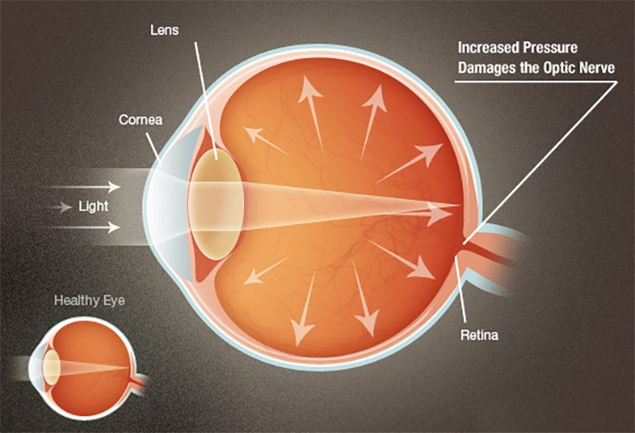
A clear fluid called the ‘Aqueous Humor’ circulates continuously within the eye. This fluid is not a part of the tears on the outer surface of the eye. Produced behind the Iris (The brown part of the eye), it flows forwards through the pupil and drains out of the eye through intricate drainage channels. Normally, fluid production and outflow are balanced. As a result the pressure inside the eye remains stable and within the safe range. If the drainage of this fluid is hampered, the pressure within the eye increases to a level that damages the cells in the optic nerve. This is Glaucoma (Kala Motia).


About Rise in pressure
The drainage portion of the eye, called the ‘drainage angle’ is like a sieve and can be blocked in different ways.
a) It may get blocked suddenly by the iris that closes the drainage angle. Eye pressure increases rapidly, resulting in a sudden loss of vision, severe eye pain and headache, rainbow halos around light accompanied by nausea and vomiting. This is called “Acute Angle Closure Glaucoma” and if not treated as an emergency it leads to permanent blindness.
b) In the second type of Glaucoma, the out flow sieves get blocked by debris. This leads to a slow rise in pressure, known as ‘Primary Open Angle Glaucoma’. Vision is lost so gradually and painlessly that a person is unaware of until the optic nerve is badly damaged. What makes it dangerous is that has no symptoms. This type of Glaucoma is much more common.
c) Glaucoma can also occur after an injury, inflammation of eye, drugs, cataract etc. Glaucoma may rarely be present at birth. The parents may notice their baby’s eye enlarging due to the increase in pressure. The cornea becomes cloudy, with watering and increasing sensitivity to light. This needs an urgent eye examination .


Range of Normal Pressure
The average Intra-Ocular Pressure (IOP; the pressure within the eye like the air pressure in tyre of a car) in adult is 16 mmHg. The actual upper limit of normal pressure, however, is difficult to pinpoint. If the 10P is consistently above 21mmHg, the chances of eye damage are probably around 10%. When the 10P is above 26mmHg, the likelihood increases to about 50%. What constitutes normal 10P is an individual matter for each person. For example, some persons with an 10P of 16mmHg may need surgery while, others with an 10P of 30mmHg may be kept under observation only. The 10P is different and independent of blood pressure. Don’t confuse it with the pressures of day to day life!


Common in all type of Glaucoma
All types of Glaucoma have three features in common. These form the basis to diagnose Glaucoma, measure the extent of damage, and monitor its progression. a) Increased 10P: The pressure inside the eye is measured with Goldmann applanation tonometer. A prism with blue lights touches the eye to accurately check 10P. There are however some types of glaucoma where damage may occur even with a normal pressure, called Normal Tension Glaucoma. b) Cupping and atrophy of the Optic Nerve: It is the drying up of the optic nerve (the nerve of sight) as it suffers damage due to high pressure inside the eye. It is assessed by examination of the fundus of the eye. c) Retinal Nevre fibre layer (RNFL) defects and Visual Field Defects: The slow death of nerve fibers is the earliest change to occur in Glaucoma. This nerve fibre layer damage is picked by a specialized instrument called OCT. Visual Fields defects are missing areas in the field of sight, though the person may be seeing well otherwise. This is measured with an instrument called a perimeter. The modern perimeter is computerized to measure; analyse, compare and report the defects.


Symptom
Unfortunately there are no symptoms in early stages. A person with Chronic Glaucoma is usually unaware of the disease. Like the hands of a clock, Chronic Glaucoma moves so slowly that its progress is not noticed. It is a silent thief of vision.
On the other hand, Acute Glaucoma, in which the pressure rises rapidly, causes severe symptoms which compel the patient to consult a doctor. Symptoms that suggest the presence of Chronic or Acute Glaucoma include are shown in above table.


Early Detection
Getting an annual eye examination at Thind Eye Hospital is the best way to detect Glaucoma. During a complete work up for Glaucoma, we will be asure your Int ra-Ocular Pres sure (Tonometry), the central corneal thickness (Pachymetry) inspect the drainage angle of the eye (Gonioscopy), evaluate for optic nerve head damage (Ophthalmoscopy), test the visual field of each eye (Perimetry) and measure the thickness of your retinal nerve fibre layer (OCT examination).
All of these tests may not be necessary for every person. But it is very important that these tests be repeated on a regular basis to monitor the progress of disease and to detect glaucoma at the earliest possible stage.


Risk Factor
People with high 10P have a higher risk of developing optic nerve damage. Other important risk factors include advancing age, severe myopia (near sighted), and a family history of Glaucoma, presence of Diabetes, past injury to the eye, surgery, or history of severe anaemia or shock.
We will weigh all these factors before deciding whether you need treatment for Glaucoma or not. If your risk of developing Glaucoma is higher than normal but there is no optic nerve damage, you will be monitored periodically as a ‘Glaucoma suspect’.


Medical Treatment
Eye drops to lower your 10P are the first line of treatment. They act to decrease eye pressure either by reducing the production of fluid within the eye or by improving the outflow through the drainage angle. It is very important to use the drops regularly at the prescribed timings.

Sometimes pills may also be required. Some medicines may occasionally result in unwanted side effects. They may sting, redden the eye and cause blurring of vision or headaches. Such side effects usually disappear after a few days. Rare side effects may be changes in your heart rate and breathing. Inform us immediately if you think you are experiencing side effects it is very important to consult your ophthalmologist before starting, stopping or changing your medication.. Frequent eye examinations and tests are the only way to monitor any changes in your Glaucoma.

Laser surgery is effective for some types of Glaucoma. In a laser surgery known as trabeculoplasty used in chronic open angle Glaucoma, the drain itself is treated In another laser surgery known as Laser iridotomy used in angle-closure Glaucoma, a hole is made in their is 0 to restore the free flow of aqueous fluid. These laser surgeries help to reduce the need for eye drops and pills to control your pressure.

In advanced cases, surgery known as trabeculectomy is necessary to control Glaucoma. We use specialized instruments to create a new drainage channel for the fluid to leave the eye, thus lowering the pressure in advanced cases. If surgery fails, special Glaucoma valves can be implanted. Fortunately, glaucoma surgery today is very safe and serious complications of modern are rare. Surgery for Glaucoma would be recommended only if the medicines or lasers fail to prevent damage to the optic nerve.

Whatever may be the approach, the objective of the treatment is to lower the eye pressure to a level at which optic nerve damage does not develop or worsen. Although eye pressure is important, the condition of your optic nerve and peripheral vision are equally or more important. It is very important to understand that achieving a normal pressure does not mean that glaucoma has been cured. It only means that the treatment is effective and would help prevent further damage to your vision. However regular eye examinations are very important while you are on treatment as glaucoma behaves differently in every patient and more aggressive treatment is often needed in some patients. This would involve reducing the pressure even further since the threshold at which pressure can cause damage is different for each individual.

The success of your treatment depends entirely on the team work put in by you and the Thind Eye Hospital team. On your part is therefore important to communicate with us and to remember to keep the follow-up appointments. If you don’t, glaucoma could be stealing your sight without your knowledge.


Management
Glaucoma is a lifelong diagnosis and treatment. Strictly speaking Glaucoma cannot be cured but it can be controlled to slow or halt optic nerve damage. The key to preventing blindness from Glaucoma is early diagnosis and treatment. This is why we emphasize regular checkups to detect glaucoma, especially if you have family history of glaucoma. If glaucoma is detected at an early stage and effective treatment is started we can prevent it from causing ANY vision loss. However achieving this goal requires a lifelong commitment to treatment and regular visits to Thind Eye Hospital.


Suggested Screening

  • People with high intra-ocular pressure (IOP): Need a baseline glaucoma work up including pressure check, central corneal thickness measurement, gonioscopy, fundus examinations OCT and Perimetry. Regular follow-up visits to monitor !OP and optic nerve damage are required every six months.
  • People over the age of forty years with no previous glaucoma: At least once every two years, especially whenever there is a change of reading glasses.
  • Family history of Glaucoma: Regular screening form a young age for people with parents or close relatives suffering from glaucoma.
  • People having high minus or plus numbered glasses: Once a year checkup.
  • Other high risk factors like diabetes, eye injuries, a major eye surgery, prolonged steroid use etc., need more frequent screening

Retinal Detachment

What is Retina ?

The delicate inner lining of the eye which is around 1 millimeter thick is known as the retina. A number of sight-threatening problems can occur in the retina. If there is any damage caused to the retina, it is possible that a patient may develop some amount of vision loss. Diabetic retinopathy, retinal tears, macular degeneration and retinal detachment are the common problems associated with retina. Any one of these conditions can result in total blindness.

Retinal detachment is a condition in which the Retina separates from the underlying layer and hence loses its functional capacity. A retinal detachment, leads to a sudden and severe loss of vision, and if not treated properly can cause blindness and shrinking and disfiguring of the eye.


Most commonly, retinal detachment occurs due to a hole/tear formation in the retina. Through this hole, liquefied vitreous, which is jelly like substance filling the eyeball, passes behind the retina and detaches it. 

Tractional retinal detachment usually occurs in diabetics or other vascular diseases of the retina. Extensive membranes are formed on the retina, which on contracting, pull the retina up.

Squint

What is Squint

Squint is misalignment of eye where two eyes are pointed in different directions. Though it is a common condition seen among 4 of 100 children it can appear in adults. The deviation can be in any directions- inwards, outwards, upwards or downwards. The misalignment may be permanent, noticeable always or it may be temporary, seen occasionally.

Causes of Squint?
Squint may be caused by any of the following:

  • Weakened muscles or abnormal nerve impulses to the eye muscles
  • Heredity
  • Blurred or poor vision ‘Conditions inside the eye such as cataract


Symptoms of squint
Main complaint of patient is that the eyes are not straight. Sometimes, a youngster will squint or close one eye in bright sunlight. Faulty depth perception may be present. Some children turn their face or tilt head in a specific direction in order to use their eyes together.


Treatment
Patient often gets the false impression that child may “outgrow” the problem. If a child has two eyes pointed in different directions, eye examination by ophthalmologist is necessary to determine the cause and to begin treatment. The goals of treatment are to preserve vision, straighten the eyes and to restore binocular vision. Treatment of squint depends upon the exact cause of the misaligned eyes. It can be directed towards unbalanced muscles or other conditions, which are causing the eyes to point in two different directions. After a complete eye check-up, including detailed study of the inner parts of the eye, an ophthalmologist can recommend appropriate optical, medical and surgical therapy.


Non Surgical Treatment
Some squints are caused because of refractive errors. For such cases, wearing prescribed glasses can correct squint completely.


Surgical Treatment
Most patients require surgical corrections. Surgery is done
under anaesthesia, in children and under local anaesthesia in adults.


Facts about Squint Surgery

  • Squint involves both eye, It may be obvious only in one eye
  • Surgery is done on eye muscles situated out side the eyeball.
  • Muscles are adjusted to correct the position of eye in straight gaze
  • One or more muscles may be operated depending upon type and severity of squint
  • Surgery is done either on both eyes simultaneously or one eye at a time
  • Squint surgery can be done in any eye
  • Other eye surgery may be required later
  • Surgery does not worsen the patients sight
  • Most of the surgeries are for “cosmetic” reasons
  • Patient may experience double vision temporally after surgery. This usually resolve in few days. Personalize Your Vision – We Make Great Vision Affordable
  • Patient should be free of any general illness
  • Parents should inform doctors of systemic disease especially cardiac problems and fits
  • Routine blood investigations and X-rays are required before surgery
  • Patient undergoing general anaesthesia should not consume water or solids foods at least four hours before surgery.
  • Anaesthetist fitness should be obtained before receiving anaesthesia
  • Eye is bandaged for one day after surgery.
  • Patients are usually freed from hospital same day when the effect of anaesthesia wears off
  • Eye drops are used for one month after surgery
  • Oral medicines are used for first few days after surgery
  • First follow—up is on next day, after one week and after one month of surgery.
  • Further follow-up depends upon patients requirement for additional treatments for Amblyopia or refractive errors etc.
  • Patients can start normal activities form next day of surgery
  • Patient should avoid water entering the eyes for at least a week after surgery
  •  

In case of doubt please consult our doctors. Child with squint do not outgrow the condition child with squint usually has defective vision which can be corrected by proper treatment if initiated early in life treatment for squint may be non-surgical which include glasses or patching of eyes surgical treatment of eye if indicated should be at the earliest possible age; this allows better surgical results and binocular function.

iLasik-Refractive Surgery

" ILASIK " AMO Innovation At Every Step

In iLasik, no blade is used to cut tissues. A hinged Corneal flap is made with the help of a laser i.e., ” Blade Free” Creation of flap. There is no cutting of tissues. They are separated by placement of the tiny bubbles in the tissues. During flap Creation the procedure can be stopped any time and restarted at a different depth without affecting the results of surgery.

Every parameter of the flap can be adjusted so as to have the best possible customized flap for that eye. Advantage of ” Blade Free “flap is that flap is very stable, well fitted and the complications encountered with the use of blade are not seen with intralase.

Over 17 + Million procedure done world wide and people found this much comfortable then any older classic spectacle removal surgery.


“ iLasik is the only approved procedure for US Air Force and Naval Pilots”


You are eligible for LASIK if…

  • You are above the age of 18 years. There is no upper limit for the procedure.
  • The power of the spectacle is equal to or more than ±0.75D.
  • A stable power of glasses that has not fluctuated by more than half a number during the previous year.


NORMAL VISION
In normal eye, in order to see clearly, rays of Light pass through the Cornea, pupil and lens of the eye . A sharp and clear image is focused directly on the retina.


MYOPIA (Near sightedness)
In a nearsighted Person the cornea is too steep or the eye is too long and the focal point is in front of the retina resulting in blurred distance vision.


HYPEROPIA (Far sightedness)
In a farsighted person the focus of the eye is Behind the retina. People who are farsighted can have trouble seeing at distance and near.


ASTIGMAGTISM
Astigmatism occurs when the cornea is not Completely round but Shaped more like an egg. Light that enters the eye focuses on more than one spot on the retina resulting in blurring of vision. Astigmatism can occur with nearsightedness or farsightedness.


LASIK surgery is a three step technique :

Step 1 – Using an instrument Called Microkeratome, a thin flap of corneal tissue is made and folded back.
Step 2 – You will be asked to look at a target light white the Excimer laser reshapes the corneal tissue.
Step 3 – The corneal flap is then placed back into its original position and allowed to dry.


Advantages of LASIK

  • Fast visual recovery
  • Minimal side effects
  • Minimal or absence of pain
  • Faster healing
  • Safest form of Refractive Surgery

Refractive Errors

What is a Refractive Error ?

Refractive error means that the shape of your eye does not bend light correctly, resulting in a blurred image. The main types of refractive errors are myopia (nearsightedness), hyperopia (farsightedness), presbyopia (loss of near vision with age), and astigmatism.

Symptoms:
  • Blurred Vision
  • Difficulty reading or seeing up close
  • Crossing of the eyes in children (esotropia)

Causes :
Overuse of the eyes does not cause or worsen refractive error. The causes of the main types of refractive error are described below:


Myopia(close objects are clear, and distant objects are blurry)
Also known as nearsightedness, myopia is usually inherited and often discovered in childhood. Myopia often progresses throughout the teenage years when the body is growing rapidly.· Hyperopia (close objects are more blurry than distant objects)
Also known as farsightedness, hyperopia can also be inherited. Children often have hyperopia, which may lessen in adulthood. In mild hyperopia, distance vision is clear while near vision is blurry. In more advanced hyperopia, vision can be blurred at all distances.


Presbyopia (aging of the lens in the eye)
After age 40, the lens of the eye becomes more rigid and does not flex as easily. As a result, the eye loses its focusing ability and it becomes more difficult to read at close range. This normal aging process of the lens can also be combined with myopia, hyperopia or astigmatism.


Astigmatism
Astigmatism usually occurs when the front surface of the eye, the cornea, has an asymmetric curvature. Normally the cornea is smooth and equally curved in all directions, and light entering the cornea is focused equally on all planes, or in all directions. In astigmatism, the front surface of the cornea is curved more in one direction than in another. This abnormality may result in vision that is much like looking into a distorted, wavy mirror. Usually, astigmatism causes blurred vision at all distances.

Amblyopia (Lazy Eye)

Amblyopia (or lazy eye) is the most common cause of poor vision in children. Amblyopia occurs because the part of the brain that deals with vision for that eye has failed to develop normally.


The vision centre in the brain is constantly developing during the first seven to eight years of a child’s life. If the vision is interfered with in any way then the brain will start to prefer one eye over the other and the vision in the other eye will suffer. To reverse the process and correct the vision in the poor eye, it is necessary to make the brain use this eye again.

Causes of Amblyopia
The most common problem is poor focusing due to myopia (nearsightedness), hyperopia (farsightedness), or astigmatism (irregularity of the focus) and Anisometropia.


Strabismus (a squint or turning eye)
Strabismus will sometimes cause amblyopia. If, for example, there is an in-turning of the eye, and it is always the same eye that turns in, then this eye will become amblyopic.


Visual deprivation
Anything that interrupts the passage of light into the eye, such as a scar on the cornea (surface of the eye), a cataract (an opaque lens in the eye), or a very droopy eyelid can lead to amblyopia.


Treating amblyopia
Treating amblyopia relies on making a child use the poorer eye in order to build the vision up again. Patching is the mainstay of amblyopia treatment. Amblyopia treatment only treats the vision and does not make a turned eye become straight.

Patching

Patching or covering the good eye is the mainstay of amblyopia treatment. Patching can be done full-time with the patch worn all day every day for a set period or it can be done part time for a certain number of hours a day, depending on the degree of amblyopia. It is easier to treat amblyopia successfully if the treatment is started while the child is young. Beyond the age of five years it becomes increasingly difficult to reverse amblyopia.
Beyond seven years it is particularly difficult. Patching to treat left eye amblyopia

Patching is very hard work for both parents and children. Most children, even in infancy, object to the patch or sometimes simply fall asleep when it is put on.

Tips on patching

Strategies for keeping the patch on: If your child is old enough to understand the reason for the patch, then often a simple explanation for why it is necessary will help. A reward system has been found by parents to also be effective. Providing interesting and supervised activities can also act to distract the child from the patch. If your child continues to struggle with patching, speak to your orthoptist who can give you information about other strategies that may be tried.

Treat skin irritation early: Some children will experience skin irritation where the patch is attached to the face. This may be due to a minor allergy to the adhesive. Switching tape/patch brands may help eliminate the problem.


Micropore tape or commercial patches: 

Usually 5cm Micropore tape is recommended for patching and our orthoptists will be able to provide instructions and tips and tricks for its use. Micropore and commercial patches are available for purchase from Thind Medical Shop and can be obtained from some chemists.


Wearing glasses:

If your child wears glasses then a patch over the spectacle lens is sometimes useful. The patch has to extend back to the forehead from the top of the glasses and along the side of the frame to ensure that child cannot see around it. Fabric patches, called opticlude, are available for purchase from Thind Medical Shop.


Alternatives to patching


Eye drops:

Atropine eye drops can be instilled on a regular basis into the good eye to blur the vision. The drops can be used on their own or in conjunction with glasses and patching. These drops act by relaxing the focusing system of the eye. They also dilate the pupil and can make the eye light sensitive. These drops will work only for certain degrees of amblyopia as they rely on blurring the good eye enough to make it worse than the amblyopic one. The drops can be given every day or just twice a week depending on your child’s eyesight. Don’t give up too soon : If the treatment is proving impossible then it is reasonable to have some time out for a few weeks before trying again. As long as the child is still young there should be time to reverse the amblyopia. There are occasionally times when amblyopia treatment doesn’t work and you may have to accept that one eye will always be poorer than the other, but it is always reassuring to know that you have done everything possible to treat it.


Glasses for Infants & Children

Glasses are prescribed for children to improve vision, prevent and treat amblyopia (‘lazy eye’), or to correct eye muscle problems. More specifcally, glasses may be required for:


1. Myopia (nearsightedness) Light rays entering the eye focus in front of the receptor cells in the retina. Myopes may see clearly at near but are blurred at distance.


2. Hyperopia (farsightedness) Light rays entering the eye focus behind the retina. The eye has to exert extra focusing power to see clearly at distance and even more so at near. If the hyperopia is large then the eye may young children have mild degrees of hyperopia.


3. Astigmatism Light rays entering the eye focus at different places. It is caused by an irregular surface of the eye. Instead of being perfectly round, the surface of the eye is shaped like a rugby ball lying on its side and is more curved in one plane than the other.


4. Anisometropia Each eye has a different focus and the worse eye may become lazy. In this situation the glasses treat the difference between the two eyes.


5. Refractive Esotropia There is a link between the focusing muscle in the eye and the muscles, which draw the eyes together when looking at a near object. Some children with hyperopia are required to exert so much focusing power in their eyes that their eyes turn in. This refractive esotropia may be fully correctable with glasses.


How are young children tested for glasses?

A retinoscope is used to shine a beam of light into the eye. Movement of the light gives a characteristic refection in the pupil indicating hyperopia, myopia or astigmatism. Lenses of varying power are placed in front of the eye until the refractive (focusing) error is cancelled out. In young children it is necessary to dilate the pupil and relax the focusing muscle with eye drops to get an accurate test. The eye drops make the child a little light-sensitive and blur the vision for several hours. The drops take 40 minutes to work fully. How are glasses prescribed? A prescription is given for each eye. The higher the first number on the prescription, the greater the correction required in the lens. If there is a second number then this means there is some astigmatism and the higher the number the greater the astigmatism. A plus sign in front of the first number indicates a hyperopic (farsighted) prescription while a minus sign indicates a myopic (nearsighted) prescription. The prescription is taken to an optometrist or optician who will prepare the lenses.


What type of lenses are the best?

Glass lenses should be avoided in children because they are more likely to break. Plastic or polycarbonate lenses are lighter and safer but they scratch more easily. The lenses must be made with a scratch-resistant hard coating. Some high power prescriptions can result in thick lenses and your optometrist or optician may advise you on modifying a lens to make it more cosmetically acceptable. High density/thinner materials can be used and the edges of the lenses can be thinned to improve the appearance.


What kind of frames are suitable for children?

The frame you choose for your child should be comfortable, safe, sturdy, and attractive. Whenever possible, purchase the glasses from an optometrist interested in working with children, and ask for a recommendation on the most suitable frame style for your child’s facial features, age,
de prescription power, and activities. Enquire about a frame guarantee, as these may be available on certain frames. Some frames can be fitted with clip-on sunglasses. They are not essential, but sunglasses are advisable for all children on bright days. Normal adult ear pieces are usually unsuitable for children and adjustments should be made so the glasses will sit more securely. For infants, straps may need to be substituted for ear-pieces to help keep the glasses in place. Flexible hinges are advisable as children tend to be careless when removing their glasses and fexible hinges tolerate a lot more abuse. If the child is old enough, let him/her help select the frame but follow your optometrist’s advice about size and fit as a child will not wear uncomfortable glasses.


How can I keep glasses on my child?

It is most important that parents are positive about the glasses. They should not make a big fuss about them but they must encourage the child to believe that the glasses suit him/her. If your child is an infant or toddler, distract him/her after you have put on the glasses. If your child removes them then replace them immediately. If the child removes them again, then put them aside for a short time and then try again. You must be patient but persistent in having the child wear the glasses.


Should my child wear glasses all the time?

Usually when a child is prescribed glasses it is because the child needs to wear them all the time. Some children have amblyopia (lazy eye’) with poor development of the visual pathways to the brain because the image into the eye has been blurred. In this situation it is essential that the child wears the glasses all the time so that the brain can learn to recognise clear images. If one eye is more long or short-sighted than the other, and sees more poorly even with the glasses then the better eye may need to be patched (covered up) for part of each day in order to build the vision in the lazy eye.


How do I care for my child’s glasses?

Children should be taught to remove their glasses using both hands without twisting the frame. They should keep them in a protective case and not put them face down on any surface. Use water or liquid soap and a soft cloth to clean them and avoid rough paper towels or tissues. If the glasses are damaged or badly scratched take them back to your optometrist for repair or replacement. Children’s glasses usually need to be readjusted every few months; more frequently than for adults.

How Does Eye Work?

The best way to understand the functioning of an eye is to compare it to a camera. Just like a camera creates images by focusing on an object and allowing specific amounts of light to pass through its aperture to create a visual impression on the film, the eye functions in the same manner.


When light enters the eye, it passes through the cornea which transmits the light onto the pupil. The pupil, similar to the aperture in a camera, adjusts the amount of light that is transmitted onto the lens. The natural lens (similar to the camera lens) alters its shape by adjusting the eye muscles in order to precisely focus the light rays onto the retina.


The retina may be equated to a camera film. It is in the retina where the image is converted into electrical signals, which are then sent to the brain. The brain interprets these signals into what we perceive as sight.

Common Eye Disorders and Diseases

More than 4.2 million Americans aged 40 years and older are either legally blind (having best-corrected visual acuity of 6/60 or worse (=20/200) in the better-seeing eye) or are with low vision (having best-corrected visual acuity less than 6/12 (<20/40) in the better-seeing eye, excluding those who were categorized as being blind).

Thind Eye Hospital Jalandhar celebrates its 32nd yrs of glorious service

Thind Eye Hospital Jalandhar celebrates its 32nd yrs of glorious service

Today Thind Eye Hospital, Jalandhar celebrates its 25th yrs of glorious service in the field of advanced Ophthalmology to the patients of Punjab, surrounding states & the NRI’s from various countries. Thind Eye Hospital popularly known as “ THIND DE JANA AE” has always remained committed to reinvest in acquisition of latest technology & machines from the world over so that State of the Art treatment available anywhere in the world can be provided to their valued patients at their door steps. Hospital has also remained equally focused to enroll highly qualified Doctors, Technicians & the staff to operate the latest technology & the machines held in their hospital.
 

Dr. Thind always had equal concern for providing latest inhouse infrastructure for the patients at reception, waiting hall at various floors & the modular theatres so that alongside the best treatment, best internal ambiance which soothens the patients & their attendants can be given.


Basic foundation of Thind Eye Hospital has been on the basis of service with humility, courtesy and the smile toward each and every patient and their attendants. The hallmark of the hospital lies that they have never differentiated in their patients irrespective of their financial status or the areas from where they hail. The hospital has very soft corner for the ex-servicemen (veterans) for their valuable services towards our nation. Thind Eye Hospital has remained committed in their future advancement and planning, keeping in view of their motto “ WE LEAD TODAY FOR TOMORROW”.


A 4 year boy’s surgery is successful.

This 4-Year-old child was rushed to Thind Eye Hospital with a 3-inch (7 cm) long nail accidentally penetrated (Head inside) in his right eye. The child was examined and advised for an X-ray to determine the exact position of the nail.


The Child was immediately taken for surgery by the doctors of the Thind Eye Hospital led by Dr. Sangeet Mittal and the nail was successfully removed. The good news is that the eye as well as the vision of the child was saved. This child is now seeing normal after the surgery. 

Before Surgery

Before Surgery

New Doctors Welcomed at Thind Eye Hospital

New Doctors Welcomed at Thind Eye Hospital